Recombinant Human Vitamin D-Binding Protein/VDB/Gc-globulin
| Product name: | Recombinant Human Vitamin D-Binding Protein/VDB/Gc-globulin |
| Source: | Human Cells |
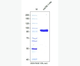
| Purity: | Greater than 95% as determined by reducing SDS-PAGE. |
| Buffer Formulation: | Lyophilized from a 0.2 μm filtered solution of 20mM PB,150mM NaCl,pH7.2. |
| Applications: | Applications:SDS-PAGE; WB; ELISA; IP. |
| Storage: | Avoid repeated freeze/thaw cycles. Store at 2-8 oC for one month. Aliquot and store at -80 oC for 12 months. |
| UOM: | 100ug/50ug/200ug/1mg/1g |
| Source | Human Cells |
| Description | Recombinant Human Vitamin D-Binding Protein is produced by our Mammalian expression system and the target gene encoding Leu17-Leu474 is expressed with a 6His tag at the C-terminus. |
| Names | Vitamin D-Binding Protein, DBP, VDB, Gc-Globulin, Group-Specific Component, GC |
| Accession # | P02774 |
| Formulation | Lyophilized from a 0.2 μm filtered solution of 20mM PB,150mM NaCl,pH7.2. |
| Shipping |
The product is shipped at ambient temperature. |
| Reconstitution |
Always centrifuge tubes before opening. Do not mix by vortex or pipetting. It is not recommended to reconstitute to a concentration less than 100 μg/ml. Dissolve the lyophilized protein in ddH2O. Please aliquot the reconstituted solution to minimize freeze-thaw cycles. |
| Storage |
Lyophilized protein should be stored at < -20°C, though stable at room temperature for 3 weeks. Reconstituted protein solution can be stored at 4-7°C for 2-7 days. Aliquots of reconstituted samples are stable at < -20°C for 3 months. |
| Purity |
Greater than 95% as determined by reducing SDS-PAGE. |
| Endotoxin | Less than 0.1 ng/µg (1 IEU/µg) as determined by LAL test. |
| Amino Acid Sequence |
LERGRDYEKNKVCKEFSHLGKEDFTSLSLVLYSRKFPSGTFEQVSQLVKEVVSLTEACCAEGADP DCYDTRTSALSAKSCESNSPFPVHPGTAECCTKEGLERKLCMAALKHQPQEFPTYVEPTNDEICE AFRKDPKEYANQFMWEYSTNYGQAPLSLLVSYTKSYLSMVGSCCTSASPTVCFLKERLQLKHLSL LTTLSNRVCSQYAAYGEKKSRLSNLIKLAQKVPTADLEDVLPLAEDITNILSKCCESASEDCMAK ELPEHTVKLCDNLSTKNSKFEDCCQEKTAMDVFVCTYFMPAAQLPELPDVELPTNKDVCDPGNTK VMDKYTFELSRRTHLPEVFLSKVLEPTLKSLGECCDVEDSTTCFNAKGPLLKKELSSFIDKGQEL CADYSENTFTEYKKKLAERLKAKLPDATPTELAKLVNKRSDFASNCCSINSPPLYCDSEIDAELK NILVDHHHHHH
|
| Background | Vitamin D-Binding Protein (DBP) is a member of the ALB/AFP/VDB family. DBP is a secreted protein and contains three albumin domains. The primary structure contains 28 cysteine residues forming multiple disulfide bonds. DBP acts as a multifunctional protein found in plasma, ascitic fluid, cerebrospinal fluid, and urine and on the surface of many cell types. DBP binds to vitamin D and its plasma metabolites and transports them to target tissues. DBP associates with membrane-bound immunoglobulin on the surface of B-lymphocytes and with IgG Fc receptor on the membranes of T-lymphocytes. |


