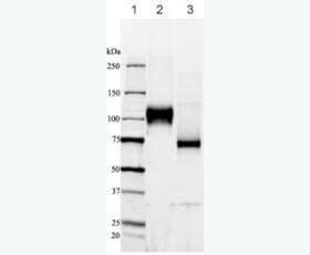
Recombinant Human Vascular Cell Adhesion Protein 1/VCAM-1/CD106/L1CAM
| Product name: | Recombinant Human Vascular Cell Adhesion Protein 1/VCAM-1/CD106/L1CAM |
| Source: | Human Cells |
| Purity: | Greater than 95% as determined by reducing SDS-PAGE. |
| Buffer Formulation: | Lyophilized from a 0.2 μm filtered solution of 20mM PB, 150mM NaCl, 2mM CaCl2, 2mM MgCl2, 5% Threhalose, pH 7.2. |
| Applications: | Applications:SDS-PAGE; WB; ELISA; IP. |
| Storage: | Avoid repeated freeze/thaw cycles. Store at 2-8 oC for one month. Aliquot and store at -80 oC for 12 months. |
| UOM: | 100ug/50ug/200ug/1mg/1g |
| Source | Human Cells |
| Description | Recombinant Human Vascular Cell Adhesion Protein 1 is produced by our Mammalian expression system and the target gene encoding Phe25-Glu698 is expressed with a 6His tag at the C-terminus. |
| Names | Vascular Cell Adhesion Protein 1, V-CAM 1, VCAM-1, INCAM-100, CD106, VCAM1, L1CAM |
| Accession # | P19320 |
| Formulation | Lyophilized from a 0.2 μm filtered solution of 20mM PB, 150mM NaCl, 2mM CaCl2, 2mM MgCl2, 5% Threhalose, pH 7.2. |
| Shipping |
The product is shipped at ambient temperature. |
| Reconstitution |
Always centrifuge tubes before opening. Do not mix by vortex or pipetting. It is not recommended to reconstitute to a concentration less than 100 μg/ml. Dissolve the lyophilized protein in ddH2O. Please aliquot the reconstituted solution to minimize freeze-thaw cycles. |
| Storage |
Lyophilized protein should be stored at < -20°C, though stable at room temperature for 3 weeks. Reconstituted protein solution can be stored at 4-7°C for 2-7 days. Aliquots of reconstituted samples are stable at < -20°C for 3 months. |
| Purity |
Greater than 95% as determined by reducing SDS-PAGE. |
| Endotoxin | Less than 0.1 ng/µg (1 IEU/µg) as determined by LAL test. |
| Amino Acid Sequence |
FKIETTPESRYLAQIGDSVSLTCSTTGCESPFFSWRTQIDSPLNGKVTNEGTTSTLTMNPVSFGN EHSYLCTATCESRKLEKGIQVEIYSFPKDPEIHLSGPLEAGKPITVKCSVADVYPFDRLEIDLLK GDHLMKSQEFLEDADRKSLETKSLEVTFTPVIEDIGKVLVCRAKLHIDEMDSVPTVRQAVKELQV YISPKNTVISVNPSTKLQEGGSVTMTCSSEGLPAPEIFWSKKLDNGNLQHLSGNATLTLIAMRME DSGIYVCEGVNLIGKNRKEVELIVQEKPFTVEISPGPRIAAQIGDSVMLTCSVMGCESPSFSWRT QIDSPLSGKVRSEGTNSTLTLSPVSFENEHSYLCTVTCGHKKLEKGIQVELYSFPRDPEIEMSGG LVNGSSVTVSCKVPSVYPLDRLEIELLKGETILENIEFLEDTDMKSLENKSLEMTFIPTIEDTGK ALVCQAKLHIDDMEFEPKQRQSTQTLYVNVAPRDTTVLVSPSSILEEGSSVNMTCLSQGFPAPKI LWSRQLPNGELQPLSENATLTLISTKMEDSGVYLCEGINQAGRSRKEVELIIQVTPKDIKLTAFP SESVKEGDTVIISCTCGNVPETWIILKKKAETGDTVLKSIDGAYTIRKAQLKDAGVYECESKNKV GSQLRSLTLDVQGRENNKDYFSPEVDHHHHHH
|
| Background | VCAM-1 is a single-pass type I membrane protein, contains 7 Ig-like C2-type domains. It is an endothelial ligand for very late antigen-4 (VLA-4) and α4ß7 integrin expressed on leukocytes, and thus mediates leukocyte-endothelial cell adhesion and signal transduction. VCAM-1 expression is induced on endothelial cells during inflammatory bowel disease, atherosclerosis, allograft rejection, infection, and asthmatic responses. During these responses, VCAM-1 forms a scaffold for leukocyte migration. VCAM-1 also activates signals within endothelial cells resulting in the opening of an "endothelial cell gate" through which leukocytes migrate. VCAM-1 has been identified as a potential anti-inflammatory therapeutic target, the hypothesis being that reduced expression of VCAM-1 will slow the development of atherosclerosis. In addition, VCAM-1-activated signals in endothelial cells are regulated by cytokines indicating that it is important to consider both endothelial cell adhesion molecule expression and function during inflammatory processes. |


