


| ELISA kit |
| Clinical diagnosis |
| Trfia |
| ANTIBODIES |
| CLIA kits |
| Proteins |
| New Products |
| > Human |
| > Canine |
| > Bovine |
| > Equine |
| > Fish |
| > Mouse |
| > Rat |
| > Chicken |
| > Porcine |
| > Rabbit |
| > Goat |
| > Simian |
| > General |
| > Cavia |
| > Caprine |
| > Sheep |
| > Cattle |

GFP antibody
Instruction Manual!
| Product name: | GFP antibody |
| Source: | Rabbit |
| Purity: | >95% |
| Buffer Formulation: | 0.02M Potassium Phosphate, 0.15M Sodium Chloride. 0.01% Sodium Azide was added as a preservative. |
| Applications: | EMELISAICC/IFIHC-FIHC-PIPWB |
| Storage: | Keep as concentrated solution. Aliquot and store at -20ºC or below. Avoid multiple freeze-thaw cycles. |
| UOM: | 100ug |
GFP antibody
Catalog Number:IC254457
- 1
- 2
Product Profile
| Product Name | GFP antibody |
|---|---|
| Antibody Type | Tags Antibodies |
| Productdescriptio |
|
| Immunogen |
Highly purified recombinant GFP made in Escherichia coli |
Key Feature
| Clonality | Polyclonal |
|---|---|
| Isotype | IgG |
| Host Species | Rabbit |
| Tested Applications | |
|
For WB: Use at a dilution of 1:5000~1:20000.:
|
|
| Concentration | 1mg/ml |
| Purification | Affinity purified |
Target Information
| Gene Synonyms |
Green Fluorescent Protein
|
|---|---|
| Target | GFP |
| TissueSpecificity | Reactive against all variants of Aequorea victoria GFP such as S65T-GFP, RS-GFP, YFP and EGFP. |
Database Links
Application
-
Application
Western blot of GFP protein detected with anti-GFP antibody (1:1000 dilution) and detected with IRDye® 800-conjugated goat anti-rabbit IgG (1:10,000 dilution). Wild type GFP (0.1 µg) was used to spike 30 µg of a HeLa whole cell lysate. This antibody detects a 27 kDa band corresponding to the epitope tag GFP.
-

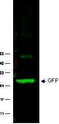
Application
Western Blot of Rabbit anti-GFP antibody . Lane 1: 293FT cells transfected with CDK4 dominant negative (C-). Lane 2: 293FT cells poitive control (C+). Load: 25 µg per lane. Primary antibody: GFP antibody at 1:400 for overnight at 4°C. Secondary antibody: Rabbit secondary antibody at 1:10,000 for 45 min at RT. Block: 5% BLOTTO overnight at 4°C. Predicted/Observed size: 27 kDa for GFP.
-

Application
Immuno-microscopy of Rabbit anti-GFP antibody . Monocyte derived dendritic cells and dermal macrophages were challenged and directly visualized with eGFP labeled Dengue virus to localize sequestration of virus particles in the different cells (upper). The location of the GFP was confirmed by TEM (lower magnified view) using GeneTex rabbit anti GFP Primary antibody (1:200) and a gold labeled secondary antibody.
-

Application
Immunofuorescence using anti-GFP antibody to label GFP-expressing glial cells (green) transplanted into lesioned rat spinal cord. Axons are labelled red by an antibody to neurofilament-200 and a rhodamine secondary. Image courtesy of Andrew Toft.
| Application Notes |
For WB: Use at a dilution of 1:5000~1:20000.:
|
|---|
Additional Information
| Form | Liquid |
|---|---|
| StorageInstructions | Keep as concentrated solution. Aliquot and store at -20ºC or below. Avoid multiple freeze-thaw cycles. |
| Storage Buffer | 0.02M Potassium Phosphate, 0.15M Sodium Chloride. 0.01% Sodium Azide was added as a preservative. |


