


| ELISA kit |
| Clinical diagnosis |
| Trfia |
| ANTIBODIES |
| CLIA kits |
| Proteins |
| New Products |
| > Human |
| > Canine |
| > Bovine |
| > Equine |
| > Fish |
| > Mouse |
| > Rat |
| > Chicken |
| > Porcine |
| > Rabbit |
| > Goat |
| > Simian |
| > General |
| > Cavia |
| > Caprine |
| > Sheep |
| > Cattle |

GFP antibody
Instruction Manual!
| Product name: | GFP antibody |
| Source: | Rabbit |
| Purity: | >95% |
| Buffer Formulation: | Keep as concentrated solution. Aliquot and store at -20ºC or below. Avoid multiple freeze-thaw cycles. |
| Applications: | ELISAICC/IFIHCIHC-FIHC-PIPWBIHC-W |
| Storage: | 1XPBS, 20% Glycerol (pH7). 0.025% ProClin 300 was added as a preservative. |
| UOM: | 100ug |
GFP antibody
Catalog Number:IC254460
- 1
- 2
- 3
Product Profile
| Product Name | GFP antibody |
|---|---|
| Antibody Type | Tags Antibodies |
| Product description |
|
Key Feature
| Clonality | Polyclonal |
|---|---|
| Isotype | IgG |
| Host Species | Rabbit |
| Tested Applications | |
|
*Optimal dilutions/concentrations should be determined by the researcher.
:
|
|
| Species Reactivity | |
| Concentration | 1 mg/ml (Please refer to the vial label for the specific concentration) |
| Purification | Affinity purified |
Target Information
| Gene Synonyms |
Green Fluorescent Protein
More
GFP eGFP enhanced green fluorescent protein |
|---|---|
| Molecular Weight(MW) | 29 kDa (note) |
| Tissue Specificity | This antibody reacts with GFP variants including GFP, EGFP, and YFP. |
Database Links
Application
-
Application
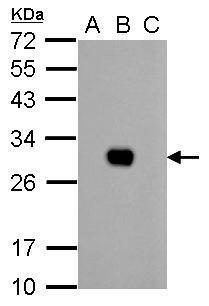
GFP antibody detects GFP protein by western blot analysis. A. 1 µg 293T whole cell extract B. 1 µg 293T whole cell extract expressing GFP-tagged protein C. 1 µg 293T whole cell extract expressing RFP-tagged protein 12% SDS-PAGE GFP antibody dilution: 1:10000 The HRP-conjugated anti-rabbit IgG antibody was used to detect the primary antibody.
-

Application
Immunohistochemical analysis (whole mount) of transgenic zebrafish embryo, using GFP antibody at 1:200 dilution.
-

Application
IP-WB assay to show that Hice1 co-immunoprecipitated with other Augmin components HAUS6 , HAUS2 and HAUS1 in U2OS cells. The HRP-conjugated anti-rabbit IgG antibody was used to detect the primary antibody.
-

Application
Immunofluorescence analysis of GFP-transfected HeLa. A:GFP is expressed in the tranfected cell. B: The cell expressing GFP can be detected using rabbit anti-GFP ab ( 1:5000) followed by Alexa Fluor 594 (1:500) goat anti-rabbit IgG. C: Merged with DNA probe, the lower cell with no GFP expressed as a negtive control.
-

Application
GFP was immunoprecipitated from bacterial GFP-expressing lysate using 1 ug of anti-GFP antibody . The precipitated GFP was detected by diluted at 1:15000.
-

Application
GFP antibody detects GFP protein by western blot analysis. Non-transfected (-) and GFP-transfected (+, ) 293T whole cell extracts (1 µg) were separated by 12% SDS-PAGE, and the membrane was blotted with GFP antibody at a dilution of 1:10000. The HRP-conjugated anti-rabbit IgG antibody was used to detect the primary antibody.
| PositiveControl | human GFP-transfected 293T , 293T whole cell extract expressing GFP-tagged protein , *GFP-p53 transfected U2OS |
|---|---|
| ApplicationNotes |
*Optimal dilutions/concentrations should be determined by the researcher.
:
|
Additional Information
| Form | Liquid |
|---|---|
| StorageInstructions | Keep as concentrated solution. Aliquot and store at -20ºC or below. Avoid multiple freeze-thaw cycles. |
| Storage Buffer | 1XPBS, 20% Glycerol (pH7). 0.025% ProClin 300 was added as a preservative. |


