Anti-rabbit IgG (H+L), F(ab') 2 Fragment (Alexa Fluor ® 555 Conjugate)
| Product name: | Anti-rabbit IgG (H+L), F(ab') 2 Fragment (Alexa Fluor ® 555 Conjugate) |
| Source: | Rabbit |
| Purity: | >95% |
| Buffer Formulation: | phosphate buffered saline , pH 7.4, 150mM NaCl, 0.02% sodium azide and 50% glycerol. |
| Applications: | WB, IP, IHC, IF, FC, ChIP |
| Storage: | The optimal dilution of the anti-species antibody should be determined for each primary antibody by titration. However, a final dilution of 1:1000 should yield acceptable results for immunofluorescent assays. Storage: Supplied in 0.1 M sodium phosphate, 0 |
| UOM: | 100ug |
Anti-rabbit IgG (H+L), F(ab') 2 Fragment (Alexa Fluor ® 555 Conjugate)
Catalog Number:IC296281
Product Profile
| Product Name | Anti-rabbit IgG (H+L), F(ab') 2 Fragment (Alexa Fluor ® 555 Conjugate) |
|---|---|
| Antibody Type | Secondary Antibodies |
| Productdescription |
|
Key Feature
| Clonality | Monoclonal |
|---|---|
| Tested Applications | |
| Species Reactivity | |
| Concentration | 1mg/ml |
| Purification |
Target Information
| TissueSpecificity |
F(ab')2 fragments are prepared from goat antibodies that have been adsorbed against pooled human serum, mouse serum, plasmacytoma/hybridoma proteins and purified human paraproteins. |
|---|
Database Links
Application
-

Application
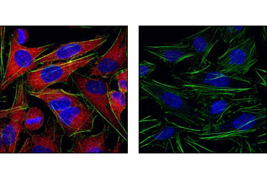
Confocal immunofluorescent analysis of HeLa cells labeled with MEK1/2 (47E6) Rabbit mAb detected with Anti-Rabbit IgG (H+L), F(ab') 2 Fragment (Alexa Fluor ® 555 Conjugate) (red, left) compared to an isotype control (right). Actin filaments have been labeled with fluorescein phalloidin (green). Blue pseudocolor = DRAQ5 ® (fluorescent DNA dye).
-

Application
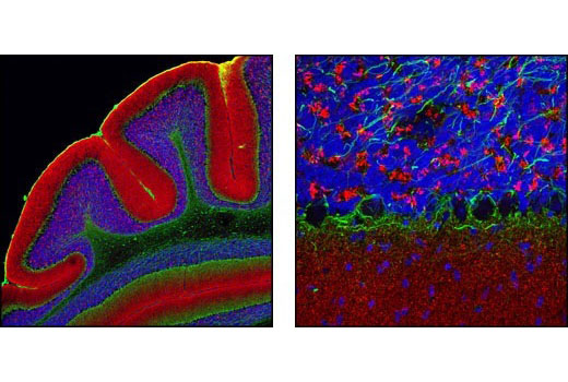
Confocal immunofluorescent analysis of mouse cerebellum using α-Synuclein Antibody (IF Preferred) detected with Anti-Rabbit IgG (H+L), F(ab') 2 Fragment (Alexa Fluor ® 555 Conjugate) (red) and Neurofilament-L (DA2) Mouse mAb detected with Anti-Mouse IgG (H+L), F(ab') 2 Fragment (Alexa Fluor ® 488 Conjugate) (green). Blue pseudocolor = DRAQ5 ® (fluorescent DNA dye).
Additional Information
| StorageInstructions |
The optimal dilution of the anti-species antibody should be determined for each primary antibody by titration. However, a final dilution of 1:1000 should yield acceptable results for immunofluorescent assays. Storage: Supplied in 0.1 M sodium phosphate, 0.1 M sodium chloride, pH 7.5, 5 mM sodium azide. Store at 4°C. Do not aliquot the antibody. Protect from light. Do not freeze. |
|---|---|
| Storage Buffer | phosphate buffered saline , pH 7.4, 150mM NaCl, 0.02% sodium azide and 50% glycerol. |


