


| ELISA kit |
| Clinical diagnosis |
| Trfia |
| ANTIBODIES |
| CLIA kits |
| Proteins |
| New Products |
| > Human |
| > Canine |
| > Bovine |
| > Equine |
| > Fish |
| > Mouse |
| > Rat |
| > Chicken |
| > Porcine |
| > Rabbit |
| > Goat |
| > Simian |
| > General |
| > Cavia |
| > Caprine |
| > Sheep |
| > Cattle |

Anti-rabbit IgG (H+L) (DyLight™ 680 Conjugate)
Instruction Manual!
| Product name: | Anti-rabbit IgG (H+L) (DyLight™ 680 Conjugate) |
| Source: | Rabbit |
| Purity: | >95% |
| Buffer Formulation: | phosphate buffered saline , pH 7.4, 150mM NaCl, 0.02% sodium azide and 50% glycerol. |
| Applications: | WB |
| Storage: | The optimal dilution of the anti-species antibody should be determined by the user. However, the final dilutions below should yield acceptable results for the respective applications. Fluorescent western blotting: 1:15000 In-Cell Western: 1:500 Storage: S |
| UOM: | 100ug |
Anti-rabbit IgG (H+L) (DyLight™ 680 Conjugate)
Catalog Number:IC296285
Product Profile
| Product Name | Anti-rabbit IgG (H+L) (DyLight™ 680 Conjugate) |
|---|---|
| Antibody Type | Secondary Antibodies |
| Productdescription |
|
Key Feature
| Clonality | Monoclonal |
|---|---|
| Tested Applications | |
| Species Reactivity | |
| Concentration | 1mg/ml |
| Purification |
Target Information
| TissueSpecificity |
Anti-rabbit IgG (H+L) (DyLight™ 680 Conjugate) reacts with heavy and light chain of most rabbit immunoglobulins. No cross-reactivity to other serum proteins has been detected. This antibody may cross-react with immunoglobulins from other species. |
|---|
Database Links
Application
-
Application
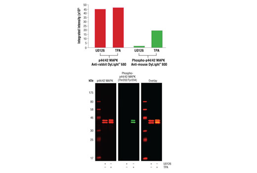
Western blot analysis of Jurkat cell lysates (#9194) treated with either U0126 (MEK 1/2 inhibitor) or TPA (12-O-Tetradecanoylphorbol-13-Acetate) using p44/42 MAPK (Erk1/2) (137F5) Rabbit mAb detected with Anti-rabbit IgG (H+L) (DyLight™ 680 Conjugate) (red) and Phospho-p44/42 MAPK (Erk1/2) (Thr202/Tyr204) (E10) Mouse mAb detected with Anti-mouse IgG (H+L) (DyLight™ 800 Conjugate) (green). The array image pixel intensities obtained using a LI-COR ® Biosciences Odyssey ® Infrared Imaging System are shown in the upper panel while corresponding fluorescent western blots are shown in the lower panel.
-

Application
In-Cell Western™ analysis of A549 cells exposed to varying concentrations of U0126 (MEK1/2 Inhibitor) for 3 hours, followed by TPA (Phorbol-12-Myristate-13-Acetate) stimulation for 30 minutes. With increasing concentrations of U0126, a significant decrease (~5 fold) in Phospho-p44/42 MAPK (Erk1/2) (Thr202/Tyr204) (D13.14.4E) XP ® Rabbit mAb signal as compared to the TPA-stimulated control was observed. When using phospho-Erk as a measurement, the IC 50 of this compound was 1.9 μM. Data and images were generated on the LI-COR ® Biosciences Odyssey ® Infrared Imaging System using Anti-rabbit IgG (H+L) (DyLight™ 680 Conjugate).
Additional Information
| StorageInstructions |
The optimal dilution of the anti-species antibody should be determined by the user. However, the final dilutions below should yield acceptable results for the respective applications. Fluorescent western blotting: 1:15000 In-Cell Western: 1:500 Storage: Supplied in 100 mM PBS, pH 7.2, containing 1% BSA and 0.02% sodium azide. Store at 4°C. Protect from light. Do not freeze. |
|---|---|
| Storage Buffer | phosphate buffered saline , pH 7.4, 150mM NaCl, 0.02% sodium azide and 50% glycerol. |


